
近日,美国麻省理工学院So, Peter T. C.团队研究了脑细胞内NADH的多光子、无标记光声和光学成像。相关论文于2025年8月7日发表在《光:科学与应用》杂志上。
在大脑中以单细胞分辨率对生物事件进行无标记检测,可以无创地捕获大脑状态,用于医学诊断和基础神经科学研究。NADH是一种通用的辅酶,不仅在细胞代谢中起着核心作用,而且可以作为捕获脑细胞和结构代谢过程的生物标志物。
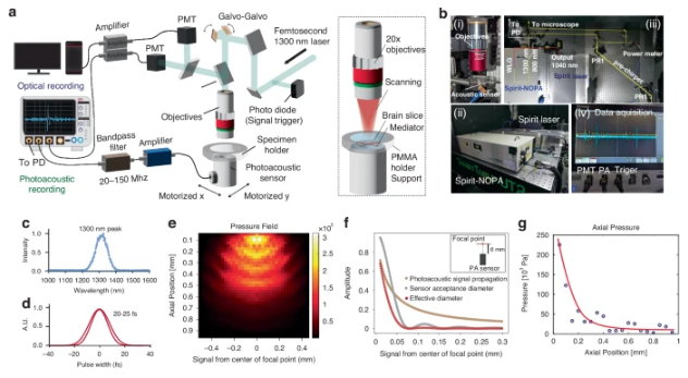
利用近红外飞秒激光,研究组研制了一种新型无标记多光子光学显微镜(LF-MP-PAM),用于观察活细胞内源性NAD(P)H。全光学方法对组织中NAD(P)H的成像深度限制在~100μm在脑组织中被近紫外荧光强吸收。低量子产率荧光团NAD(P)H的多光子(三光子)激发热特征的光学检测允许在前所未有的深度进行检测,而聚焦激发确保了高空间分辨率。
研究组通过监测在NADH溶液中培养的HEK293T细胞和HepG2细胞内NAD(P)H的增加,验证了NAD(P)H的光敏检测。他们还证明了内源性NAD(P)H光信号在700 μm深度的脑切片和1100 μm深度的脑类器官中检测到。最后,研究组开发并演示了利用实时图像采集和处理管道同时对脑细胞中的NAD(P)H进行光敏和光学成像。这种方法可以在人类和动物大脑深处的单细胞水平上,为监测大脑发育和疾病期间的代谢变化,以及神经元活动引起的变化打开一扇新的大门。
附:英文原文
Title: Multi-photon, label-free photoacoustic and optical imaging of NADH in brain cells
Author: Osaki, Tatsuya, Lee, W. David, Zhang, Xiang, Zubajlo, Rebecca E., Balcells-Camps, Mercedes, Edelman, Elazer R., Anthony, Brian W., Sur, Mriganka, So, Peter T. C.
Issue&Volume: 2025-08-07
Abstract: Label-free detection of biological events at single-cell resolution in the brain can non-invasively capture brain status for medical diagnosis and basic neuroscience research. NADH is an universal coenzyme that not only plays a central role in cellular metabolism but may also be used as a biomarker to capture metabolic processes in brain cells and structures. We have developed a new label-free, multiphoton photoacoustic microscope (LF-MP-PAM) with a near-infrared femtosecond laser to observe endogenous NAD(P)H in living cells. The imaging depth of NAD(P)H in tissues with all-optical methods is limited to ~100μm in brain tissue by the strong absorption of the near-ultraviolet fluorescence. Here, acoustic detection of the thermal signature of multi-photon (three-photon) excitation of NAD(P)H, a low quantum yield fluorophore, allows detection at an unprecedented depth while the focused excitation ensures high spatial resolution. We validated the photoacoustic detection of NAD(P)H by monitoring an increase in intracellular NAD(P)H in HEK293T cells and HepG2 cells incubated in NADH solution. We also demonstrated the detection of endogenous NAD(P)H photoacoustic signals in brain slices to 700 μm depth and in cerebral organoids to 1100 μm depth. Finally, we developed and demonstrated simultaneous photoacoustic and optical imaging of NAD(P)H in brain cells with a real-time image acquisition and processing pipeline. This approach could open a new door to monitor brain metabolic changes during development and disease, and changes due to neuronal activity, at single-cell level deep in the brains of both humans and animals.
DOI: 10.1038/s41377-025-01895-x
Source: https://www.nature.com/articles/s41377-025-01895-x
Light: Science & Applications:《光:科学与应用》,创刊于2012年。隶属于施普林格·自然出版集团,最新IF:19.4
官方网址:https://www.nature.com/lsa/
投稿链接:https://mts-lsa.nature.com/cgi-bin/main.plex