
北京大学谢晓亮研究组取得一项新突破。他们的最新研究揭示了单细胞Micro-C以高分辨率描绘3D基因组结构,并表征多增强子中心。这一研究成果于2025年7月2日发表在国际顶尖学术期刊《自然—遗传学》上。
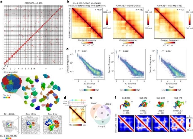
在这里,课题组研究人员报告了单细胞Micro-C (scMicro-C),一种基于微球菌核酸酶的三维(3D)基因组作图技术,其空间分辨率提高了5kb并确定了一种特殊的3D增强子结构,称为“启动子-增强子条纹(PESs)”,将基因的启动子连接到多个增强子。PES是由内聚素介导的环外离子形成的,它可能会给启动子带来多种增强子。此外,研究团队观察到,在单细胞3D基因组结构中,PES基因上的多增强子中心普遍存在,其中多个增强子与基因启动子形成一个空间簇。通过其提高的分辨率,scMicro-C阐明了增强子如何在空间上协调以控制基因。
据介绍,在动物基因组中,被称为增强子的调控DNA元件控制着特定细胞类型中精确的时空基因表达模式。然而,增强子在核仁内调节靶基因的空间组织仍然知之甚少。
附:英文原文
Title: Single-cell Micro-C profiles 3D genome structures at high resolution and characterizes multi-enhancer hubs
Author: Wu, Honggui, Zhang, Jiankun, Tan, Longzhi, Xie, Xiaoliang Sunney
Issue&Volume: 2025-07-02
Abstract: In animal genomes, regulatory DNA elements called enhancers govern precise spatiotemporal gene expression patterns in specific cell types. However, the spatial organization of enhancers within the nucleus to regulate target genes remains poorly understood. Here we report single-cell Micro-C (scMicro-C), a micrococcal nuclease-based three-dimensional (3D) genome mapping technique with an improved spatial resolution of 5kb, and identified a specialized 3D enhancer structure termed ‘promoter–enhancer stripes (PESs)’, connecting a gene’s promoter to multiple enhancers. PES are formed by cohesin-mediated loop extrusion, which potentially brings multiple enhancers to the promoter. Further, we observed the prevalence of multi-enhancer hubs on genes with PES within single-cell 3D genome structures, wherein multiple enhancers form a spatial cluster in association with the gene promoter. Through its improved resolution, scMicro-C elucidates how enhancers are spatially coordinated to control genes.
DOI: 10.1038/s41588-025-02247-6
Source: https://www.nature.com/articles/s41588-025-02247-6
Nature Genetics:《自然—遗传学》,创刊于1992年。隶属于施普林格·自然出版集团,最新IF:41.307
官方网址:https://www.nature.com/ng/
投稿链接:https://mts-ng.nature.com/cgi-bin/main.plex