|
|
|
|
|
新型光衍生质谱分析实现脂质深度结构解析|Nature Communications |
|
|
论文标题:Large-scale lipid analysis with C=C location and sn-position isomer resolving power
期刊:Nature Communications
作者:Wenbo Cao, Simin Cheng et.al
发表时间:2020/01/17
数字识别码:10.1038/s41467-019-14180-4
微信链接:点击此处阅读微信文章
脂质作为四大类生物分子和三大类营养素之一,不仅是质膜基本组成单元和能量的载体,也在信号转导和蛋白质功能调控等方面发挥着积极和关键性作用。尽管大规模脂质的质谱分析已非常成熟,但脂质的精细结构解析问题一直没有解决,严重阻碍了脂质的生物学功能研究。从结构上看,生物体内脂质的复杂性源于脂质类型、脂肪酸链及sn位置、以及碳碳双键(C=C)数量、位置及几何结构等多重结构特征。
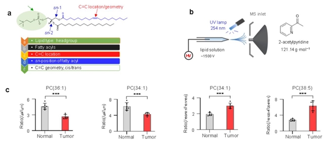
为了利用单一方法实现脂质的近全结构解析,清华大学精密仪器系质谱研究团队于近期在Nature Communications上发表了题为“Large-scale lipid analysis with C=C location and sn-position isomer resolving power”的论文。该研究基于光衍生试剂2-乙酰吡啶和串联质谱,发展了可用于大规模脂质精细结构解析的策略。该方法通过单次分析即可实现磷脂类型、脂肪酸链及sn位置、C=C位置等结构信息的全面获取,并实现脂质C=C和sn位置异构体的定量分析。该方法可在大多数具备多级质谱功能的商用质谱仪上实现,无需对质谱仪进行改造,具有广阔的应用空间。
在生物医学应用方面,利用该方法对人不同亚型的乳腺癌细胞开展了深度脂质组学分析,揭示出脂质C=C异构体组成随乳腺癌细胞侵袭性连续变化,表明脂质C=C异构体有可能作为癌症进展的标记物,后续将对此深入研究。此外,通过脂质C=C及sn两类异构体的定性和定量分析,成功地区分出II型糖尿病人血浆和人肺癌组织,表明脂质深度结构分析有用于疾病诊断的潜力。

图1: 脂质异构体的全面结构解析。a. 脂质五个层面的结构信息。b.用于脂质深度结构解析的在线光化学衍生-串联质谱方法。c.癌及癌旁组织中脂质C=C/sn位置异构体组成对比。
有奖调研
我们正在进行公众号的读者调研。您对我们的公众号有什么想法或建议?欢迎反馈给我们,帮助我们提供更好的内容和服务!
马上点击链接参与问卷调查吧!
预计完成时间:3-5分钟
抽奖奖品:“Nature”纪念版小鼠公仔 或《自然的音符》(共20份)
如果您对本次调查有任何疑问或在问卷填写过程中遇到任何问题,请发送邮件至:china@nature.com
摘要:Lipids play a pivotal role in biological processes and lipid analysis by mass spectrometry (MS) has significantly advanced lipidomic studies. While the structure specificity of lipid analysis proves to be critical for studying the biological functions of lipids, current mainstream methods for large-scale lipid analysis can only identify the lipid classes and fatty acyl chains, leaving the C=C location and sn-position unidentified. In this study, combining photochemistry and tandem MS we develop a simple but effective workflow to enable large-scale and near-complete lipid structure characterization with a powerful capability of identifying C=C location(s) and sn-position(s) simultaneously. Quantitation of lipid structure isomers at multiple levels of specificity is achieved and different subtypes of human breast cancer cells are successfully discriminated. Remarkably, human lung cancer tissues can only be distinguished from adjacent normal tissues using quantitative results of both lipid C=C location and sn-position isomers.
(来源:科学网)
特别声明:本文转载仅仅是出于传播信息的需要,并不意味着代表本网站观点或证实其内容的真实性;如其他媒体、网站或个人从本网站转载使用,须保留本网站注明的“来源”,并自负版权等法律责任;作者如果不希望被转载或者联系转载稿费等事宜,请与我们接洽。