论文标题:Paracrine effect of regulatory T cells promotes cardiomyocyte proliferation during pregnancy and after myocardial infarction
期刊:Nature Communications
作者:Serena Zacchigna, Valentina Martinelli, Silvia Moimas, Andrea Colliva, Marco Anzini, Andrea Nordio, Alessia Costa, Michael Rehman, Simone Vodret, Cristina Pierro, Giulia Colussi, Lorena Zentilin, Maria Ines Gutierrez, Ellen Dirkx, Carlin Long, Gianfranco Sinagra, David Klatzmann & Mauro Giacca
发表时间: 2018/06/26
数字识别码:10.1038/s41467-018-04908-z
原文链接:https://www.nature.com/articles/s41467-018-04908-z?utm_source=Other_website&utm_medium=Website_links&utm_content=RenLi-Nature-Nature_Comms-Cell_Biology-China&utm_campaign=NATCOMMS_USG_rlp8212_tcell_sciencenet_article_July_1st

近期《自然-通讯》发表的一项研究Paracrine effect of regulatory T cells promotes cardiomyocyte proliferation during pregnancy and after myocardial infarction分析了调节性T细胞对心肌细胞增殖能力的影响。这项小鼠研究揭示了允许胚胎心脏内心肌细胞增殖的细胞因子,并且表明相同的因子也可以促进母体心肌细胞的增殖。这些发现可能对治疗突发心脏病具有潜在意义。

图1:母体血清中的调节性T细胞促进心肌细胞增殖。
Zacchigna et al.
心肌细胞会在胚胎心脏发育期间增殖,但在出生后丧失这种能力,这是成年心脏在受损后不自发再生的主要原因之一。
意大利国际遗传工程与生物技术中心(ICGEB)的Serena Zacchigna及其同事发现,已知可使母体对胚胎产生免疫耐受的免疫细胞——调节性T细胞,也可以促进胚胎心肌细胞的增殖。此外,他们还表明这些免疫细胞也可能在怀孕期间促进母体心脏的心肌细胞增殖。在心脏疾病的小鼠模型中,在靠近小鼠心脏缺血伤口的部位注射调节性T细胞会刺激心肌细胞增殖,进而促进心脏再生反应。这被认为是调节性T细胞释放促增殖分子的结果。

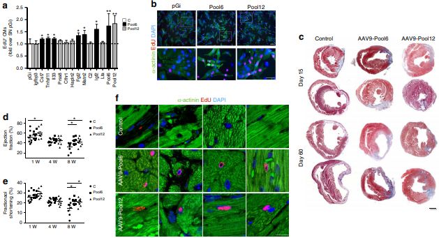
图2:调节性T细胞分泌蛋白质促进心肌细胞的增殖。
Zacchigna et al.
尽管还需要进一步的调查来证实调节性T细胞的促增殖作用,但是这些发现增加了我们对怀孕期间的心脏生物学的理解,并提出了促进心脏修复的潜在方法。(来源:科学网)
摘要:Cardiomyocyte proliferation stops at birth when the heart is no longer exposed to maternal blood and, likewise, to regulatory T cells (Tregs) that are expanded to promote maternal tolerance towards the fetus. Here, we report a role of Tregs in promoting cardiomyocyte proliferation. Treg-conditioned medium promotes cardiomyocyte proliferation, similar to the serum from pregnant animals. Proliferative cardiomyocytes are detected in the heart of pregnant mothers, and Treg depletion during pregnancy decreases both maternal and fetal cardiomyocyte proliferation. Treg depletion after myocardial infarction results in depressed cardiac function, massive inflammation, and scarce collagen deposition. In contrast, Treg injection reduces infarct size, preserves contractility, and increases the number of proliferating cardiomyocytes. The overexpression of six factors secreted by Tregs (Cst7, Tnfsf11, Il33, Fgl2, Matn2, and Igf2) reproduces the therapeutic effect. In conclusion, Tregs promote fetal and maternal cardiomyocyte proliferation in a paracrine manner and improve the outcome of myocardial infarction.
阅读论文全文请访问:https://www.nature.com/articles/s41467-018-04908-z?utm_source=Other_website&utm_medium=Website_links&utm_content=RenLi-Nature-Nature_Comms-Cell_Biology-China&utm_campaign=NATCOMMS_USG_rlp8212_tcell_sciencenet_article_July_1st
(来源:科学网)
特别声明:本文转载仅仅是出于传播信息的需要,并不意味着代表本网站观点或证实其内容的真实性;如其他媒体、网站或个人从本网站转载使用,须保留本网站注明的“来源”,并自负版权等法律责任;作者如果不希望被转载或者联系转载稿费等事宜,请与我们接洽。